
Page 29 - Demo
P. 29
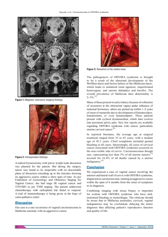
Sayyad, et al.: Carcinosarcoma in OHVIRA syndrome22 MOGS Chronicles | Volume 1 | Issue 1 | September 2024The pathogenesis of OHVIRA syndrome is thought to be a result of the abnormal development of the Wolffian ducts and fusion failure of the Mullerian ducts, which leads to unilateral renal agenesis, imperforated hemivagina, and uterine didelphys and bicollis. The overall prevalence of Mullerian duct abnormality is 2%u20133%.[1]Many of these present in early infancy because of collection of secretions in the obstructed vagina under influence of maternal hormones; others are picked up within 1%u20132 years of onset of menarche due to development of hematocolpos, hematometra, or even hematosalpinx. These patients present with cyclical dysmenorrhea, which later evolves into persistent pelvic pain. Very few reports are available regarding OHVIRA syndrome with cancer, particularly uterine cervical cancer.[2]In reported literature, the average age at surgical treatment ranged from 33 to 65 years, with a median age of 43.5 years. Chief complaints included genital bleeding in all cases. Interestingly, all cases of cervical cancer associated with OHVIRA syndrome occurred on the non-visible side of cervix. Carcinosarcomas though rare, representing less than 5% of all uterine tumors,[3]account for 16.4% of all deaths caused by a uterine malignancy.[4]ConclusionWe experienced a case of vaginal cancer involving the anterior and lateral wall of cervix with OHVIRA syndrome, diagnosed as carcinosarcoma, running an aggressive course within the span of 6 months from the onset of symptoms to its diagnosis.Combining imaging with tissue biopsy is important for women with OHVIRA syndrome who have midmenstrual bleeding or menorrhagia. The clinician should be aware that in M%u00fcllerian anomalies, cervical, vaginal malignancies may be overlooked, delaying the tumor diagnosis thus affecting patient%u2019s reproductive function and quality of life.A radical hysterectomy with pelvic lymph node dissection was planned for the patient. But during the surgery, tumor was found to be inoperable with no discernable plane of dissection extending up to the introitus showing its aggressive course within a short span of time. As per Federation of Gynecology and Obstetrics Staging for Vaginal Cancer, she had stage III vaginal cancer and T3N1MO as per TNM staging. The patient underwent chemotherapy with carboplatin but failed to respond.A trial of immunotherapy is being given in the hope of some palliative relief.DiscussionOur case is a rare occurrence of vaginal carcinosarcoma in Mullerian anomaly with an aggressive course.Figure 2: Intraoperative findingsFigure 1: Magnetic resonance imaging findingsFigure 3: Resection of the visible mass
