
Page 11 - Demo
P. 11
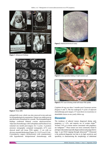
Jadkar, et al.: Management of ovarian adenocarcinoma in an IVF pregnancy4 MOGS Chronicles | Volume 1 | Issue 1 | September 2024enlarged left ovary which was also removed in toto and sent for histopathological examination. Patient was stable post op and allowed to breastfeed for a month. The histopathological findings confirmed bilateral ovarian adenocarcinoma. Adjuvant chemotherapy with Carboplatin and Paclitaxel was restarted one month post-surgery for 3 cycles. A positron emission tomography computed tomography (CT) scan showed small soft tissue FDG uptake ~2 cm with no obvious omental thickening [Figure 5]. CA125 was 8 U/mL.Cytoreductive surgery (hysterectomy with omentectomy) with hyperthermic intraperitoneal chemotherapy with Cisplatin 64 mg was done 3 months post Caesarean section [Figures 6 and 7]. She has undergone 9 cycles of adjuvant chemotherapy post-surgery. CT scan shows no recurrence or identifiable lesion at one yearly follow-up.DiscussionThe incidence of adnexal masses diagnosed during early pregnancy is 1%u20134%, and majority are of ovarian origin.[6]Around 2%u20136% of ovarian tumors associated with pregnancy are malignant.[7] These tumors are more commonly found in primigravidas and are typically diagnosed at an early stage (below stage 1c per FIGO staging) through ultrasound.[8] Ultrasound is the preferred diagnostic tool due to its high sensitivity and specificity in characterizing the morphology of abdominal Figure 1: Ultrasound findingsFigure 2: Pelvic MRIFigure 3: Adenocarcinoma of ovaryFigure 4: Intra operative finding of right ovarian cystFigure 5: PET scan showing small soft tissue FDG uptake
