
Page 17 - Demo
P. 17
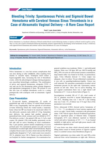
10 MOGS Chronicles | Volume 1 | Issue 1 | September 2024IntroductionPelvic hematoma is a rare but serious complication that can arise during or after childbirth, often resulting from trauma or vascular injury. Eclampsia itself creates a hypercoagulable state, increasing the risk of thrombotic events, while the physiological changes during pregnancy can exacerbate hemorrhagic conditions. The combination of pelvic and sigmoid hematomas, alongside CVST, is rare and can be life threatening unless prompt diagnosis and appropriate management is done. We present to you this rare case of multiple thrombotic event in a young primigravida with eclampsia with an atraumatic vaginal delivery.Case PresentationA 22-year-old female, primigravida, 32 weeks of gestational age with no history of raised blood pressure (BP) or any coagulopathy, had one episode of convulsion at home following which she visited nearby a maternity home where her BP was found to be raised and within 4 h she delivered a preterm male child of 1.8 kg. Immediate post delivery, the patient had another episode of generalized tonic clonic seizures and was shifted to CCU.Post delivery on day 1, the patient%u2019s Hb was 11.2 g%, total counts 12k, platelet 132k with healthy episiotomy on the left side and no active bleeding on local examination. Day 2 post partum, patient started complaining of severe perineal pain. On examination patient was pale and local examination revealed right sided vulval hematoma. Repeat complete blood count was done which showed the Hb had significantly dropped to 5.7 g%, total counts were raised to 20.3 k, platelets were 146 k. Patient was then referred to our hospital for further management. On examination her general condition was moderate, Pallor ++ and mild pedal oedema. Pulse was 130 bpm, BP was 100/70 mmHg and SPO2: 99% on room air with normal systemic examination, deep tendon reflex was found to be brisk, no premonitory signs, Urine Albumin showed +3. Urine output was adequate, clear in colour. On per abdominal examination, it was soft, no guarding/tenderness/rigidity. Uterus was well retracted. Per speculum examination revealed vulval hematoma present on the right side. Episiotomy sutures present on left side. There was no active bleeding. On per vaginal examination there was a right lateral wall hematoma of 5 %u00d7 6 cm as shown below [Figure 1].Investigations revealed, Hb of 5.7 g%, total counts of 20.3 k, platelets were146 k, lactate dehydrogenase was 941 IU/L, bleeding time was 1 min 45 s and clotting time was 5 min: 15 s, D dimer was 4.72 mg/l. Rest of the parameters were normal.Pelvic ultrasound (USG) showed pelvic hematoma of 7.1 %u00d7 5.6 %u00d7 7.1 cm adjacent to lower uterine segment and cervix on right. Uterus bulky consistent with post-partum status. USG Vulval region showed ill-defined hypoechoic fluid in superficial plane of bilateral labia. Computed tomography (CT) scan abdomen reported as bilateral vulval and pelvic hematoma of 8.9 %u00d7 5 %u00d7 9 cm located medially to right pelvic wall and right psoas muscle with oedematous sigmoid colon.CT scan brain was done which showed partial contrast filling defect noted in frontal portion of superior sagittal sinus s/o cerebral venous sinus thrombosis [Figure 2].Vaginal exploration was done under GA and 300 g clots removed from vulval hematoma on right side. However, Case ReportAbstractPelvic hematoma is a localized collection of blood outside blood vessels following trauma or injuries to blood vessels in pelvic cavity.This is a rare case of pelvic hematoma following atraumatic preterm vaginal delivery developing vulval hematoma on day 2 of delivery with sigmoid bowel hematoma and cerebral venous sinus thrombosis in a case of eclampsia.Keywords: %u200aSpontaneous pelvic hematoma, Sigmoid Hematoma, Atraumatic delivery, vulval hematomaBleeding Trinity: Spontaneous Pelvic and Sigmoid Bowel Hematoma with Cerebral Venous Sinus Thrombosis in a Case of Atraumatic Vaginal Delivery %u2013 A Rare Case ReportPreeti F. Lewis, Kumari AnkitaDepartment of Obstetrics and Gynecology, at G.G.M.C and Sir J.J. Group of Hospitals, Mumbai, Maharashtra, IndiaAddress for correspondence: Dr. Kumari Ankita, Resident, Department of Obstetrics & Gynaecology, G.G.M.C & Sir, J.J. Group of Hospitals, Mumbai, Maharashtra, India. Email: ankitamegha21@gmail.com
